ตำรายาของประเทศไทย
Thai Pharmacopoeia
สำนักยาและวัตถุเสพติด กรมวิทยาศาสตร์การแพทย์ กระทรวงสาธารณสุข
Bureau of Drug and Narcotic, Department of Medical Sciences, Ministry of Public Healthสำนักยาและวัตถุเสพติด กรมวิทยาศาสตร์การแพทย์ กระทรวงสาธารณสุข
Bureau of Drug and Narcotic, Department of Medical Sciences, Ministry of Public HealthAPPENDIX 15 ASSAY OF BIOLOGICAL PRODUCTS
15.1 HUMAN BLOOD AND BLOOD PRODUCTS
15.1.1 Determination of ABO Group of Donors
Examine the blood for both antigens on the red cells and antibodies in the serum or plasma using either manual or automated techniques.
For manual tests, collect a few ml of fresh blood without anticoagulant in a narrow test-tube, allow to clot, and remove the serum. Stir the clot or a small part of it with saline TS, centrifuge the resulting suspension, and resuspend the cells in saline TS to make a 2 to 3 per cent v/v suspension of packed cells.
For automated tests, collect a few ml of blood and prevent from clotting by the addition of 1 volume of a 10 per cent w/v solution of dipotassium edetate for each 32 volumes of blood collected. Separate the plasma and cellular components and prepare a suspension of red cells in saline TS suitable for the type of instrument to be used.
TESTS FOR ANTIGENS
Mix separate portions of the red cell suspension with Anti-A Blood-grouping Reagent, Anti-B Bloodgrouping Reagent and Anti-A,B (group O) Bloodgrouping Reagent, the specificities of which have been demonstrated by testing with red cells of known groups A, B and O, and determine the antigens present by examination of the resulting pattern of agglutination.
Manual tests Mix 1 volume of the appropriate ABO Blood-grouping Reagent with 1 volume of red cell suspension in a test-tube. Allow the tubes to stand at room temperature for 1 to 2 hours and then tap them gently to disperse the deposit of cells. Examine the contents of the tubes macroscopically for agglutination.
Automated tests Perform the tests with appropriate automatic equipment1 according to the manufacturer’s instructions. Mix the red cell suspension with the appropriate ABO Blood-grouping Reagent. Allow agglutination to take place and determine its degree either by visual inspection after the agglutinates have been deposited upon filter paper or by the use of a suitable photometric detector fitted with an automatic means of recording.
TESTS FOR ANTIBODIES
Mix separate portions of the serum or plasma with suspensions of human red cells of group A (sub-groups A1 and A2), group B and group O, and determine the antibodies present by examination of the resulting
1 Instruments based on continuous flow system or a discrete sample system may be used.
pattern of agglutination by either manual or automated tests as described above under the Tests for Antigens.
When the above tests are used to determine the blood group of an intended recipient rather than that of a donor, it should be noted that antibody development may be incomplete in infants under 1 year old; in such circumstances the blood group of the child is based solely on the antigens present on the red cells.
ABO BLOOD-GROUPING REAGENTS
(Note ABO Blood-grouping Reagents are normally selected on the basis of their suitability for use in manual tests. When such reagents are to be used in automated tests, their suitability must be reassessed by performing automated tests with a large number of red cell suspensions of different ABO group including examples of the A1, A2, A2B, and Ax sub-groups.)
ABO Blood-grouping Reagents are derived from the sera or defibrinated plasma of selected persons of the appropriate ABO blood group who may have been deliberately immunized with either red cells or groupspecific substance of the appropriate blood group or groups. Alternatively they are derived from the sera of lower animals or from cultures of mammalian lymphocytes. For preparations of human origin, use only material that has been tested with negative results for the presence of hepatitis B surface antigen, HIV antibodies, and other blood borne infectious agents by suitably sensitive methods. However, the reagent cannot be assumed to be free from infectious agents. Care must be taken in the use and disposal of each container and its contents.
ABO Blood-grouping Reagents may be issued as liquids or they may be prepared by constitution from the dried reagents. Liquid reagents are clear or faintly opalescent, yellowish or colourless fluids without turbidity. They may contain a suitable antimicrobial preservative. Dried reagents are pale yellow powders or friable solids.
ABO Blood-grouping Reagents are of three types, Anti-A Blood-grouping Reagent, Anti-B Blood-grouping Reagent and Anti-A,B (Group O) Blood-grouping Reagent.
Anti-A Blood-grouping Reagent agglutinates human red cells containing A antigens, including subgroups A1 and A2 but rarely agglutinates those red cells classified as Ax; that is, it agglutinates red cells of blood groups A and AB that include the sub-groups A1, A2, A1B, and A2B but does not agglutinate red cells of the sub-groups Ax or AxB. The reagent does not agglutinate human red cells that do not contain A antigens, i.e. red cells of blood groups O and B. The reagent is shown not to agglutinate, under the conditions specified for use, any of a comprehensive panel of group O or group B red cells chosen to bear a wide range of red cell antigens, or to contain antibodies to the serum protein factors Gm or Km. It does not agglutinate group O or group B red cells coated with IgG.
Anti-B Blood-grouping Reagent agglutinates human red cells containing B antigens, i.e. red cells of blood groups B and AB including sub-groups A1B and A2B. The reagent does not agglutinate human red cells that do not contain B antigens, i.e. red cells of blood groups O and A including sub-groups A1 and A2. The reagent is shown not to agglutinate, under the conditions specified for use, any of a comprehensive panel of group O or A red cells chosen to bear a wide range of red cell antigens, or to contain antibodies to the serum protein factors Gm or Km. It does not agglutinate group O or group A red cells coated with IgG.
Anti-A,B (Group O) Blood-grouping Reagent agglutinates human red cells containing A or B antigens; that is, it agglutinates red cells of blood groups A, B and AB including the sub-groups A1, A2, Ax, A1B, A2B, and AxB. The reagent does not agglutinate human red cells that do not contain A or B antigens, i.e. red cells of blood group O. The reagent is shown not to agglutinate, under the conditions specified for use, any of a comprehensive panel of group O cells chosen to bear a wide range of red cell antigens, or to contain antibodies to the serum protein factors Gm or Km. It does not agglutinate group O red cells coated with IgG.
ABO Blood-grouping Reagents, reconstituted where necessary as stated on the label, comply with the following requirements.
Avidity On a microscopic slide, mix a suitable volume of the reagent with an equal volume of a 5 to 10 per cent v/v suspension of human red cells of each of the groups or sub-groups given below; use a separate portion of the reagent for each type of red cell. The time taken for agglutination to appear first to the unaided eye is not more than twice that taken when the procedure is carried out using the appropriate National or International Standard of Blood-typing Serum in place of the reagent being examined.
ANTI-A BLOOD-GROUPING REAGENT Use human red cells of sub-groups A1 and A2B and preferably also use cells of sub-groups A1 and A2B; the appropriate National or International Standard is that for Anti-A bloodtyping serum.
ANTI-B BLOOD-GROUPING REAGENT Use human red cells of sub-group B; the appropriate National or International Standard is that for Anti-B blood-typing serum.
ANTI-A, B (GROUP O) BLOOD-GROUPING REAGENT Use human red cells of sub-groups A1 and A2 and of group B; the appropriate National or International Standard is that for Anti-A, B blood-typing serum.
In addition carry out the procedure described above at a temperature of 20º to 25º using red cells of subgroup Ax; agglutination first appears to the unaided eye in not more than 2 minutes.
Potency The potency of an ABO Blood-grouping Reagent is determined by comparing its “saline aggluti nin” antibody activity with that of the appropriate National or International Standard for Blood-typing serum.
The determination is carried out by simultaneously titrating the reagent being examined and the Standard against suspensions of human red cells of the groups or sub-groups given below.
ANTI-A BLOOD-GROUPING REAGENT Not less than 64 IU of anti-A antibody per ml; that is, for each type of red cell against which it is titrated, the titre of the reagent being examined is not less than one quarter of that of the Standard, irrespective of the actual titres obtained.
Use human red cells of sub-groups A1 and A2B and preferably also use cells of sub-groups A2 and A1B.
ANTI-B BLOOD-GROUPING REAGENT Not less than 64 IU of anti-B antibody per ml; that is, the titre of the reagent being examined is not less than one quarter of that of the Standard, irrespective of the actual titres obtained.
Use human red cells of group B.
ANTI-A, B (GROUP O) BLOOD-GROUPING REAGENT Not less than 64 IU of anti-A antibody and of anti-B antibody per ml; that is, the titre of reagent being examined is not less than one quarter of that of the Standard, irrespective of the actual titres obtained.
Use human red cells of sub-groups A1 and A2 and of group B.
In addition undiluted Anti-A,B Blood-grouping Reagent gives easily detectable agglutination with at least one example of red cells of sub-group Ax.
Sterility Comply with the “Sterility Test” (Appendix 10.1).
Packaging and storage ABO Blood-grouping Reagents shall be kept in sterile containers, sealed so as to exclude micro-organisms.
Liquid ABO Blood-grouping Reagents which do not contain an antimicrobial preservative shall be kept frozen, preferably at a temperature below –30º.
Liquid ABO Blood-grouping Reagents containing an antimicrobial preservative shall be kept at a temperature of 2º to 8º; they shall not be frozen unless the antimicrobial preservative has been shown to be innocuous to the reagent in the frozen state.
Dried ABO Blood-grouping Reagents shall be kept at a temperature not exceeding 20º.
Labelling The label states (1) the name “Anti-A Blood-grouping Reagent”, “Anti-B Blood-grouping Reagent” or “Anti-A,B (Group O) Blood-grouping Reagent”, as appropriate; (2) the number of IU of the relevant antibody or antibodies per ml; (3) a number or other indication by which the history of the preparation may be traced; (4) the date after which the preparation may not be expected to retain its activity; (5) the conditions under which it shall be stored; (6) for a reagent containing an antimicrobial preservative the name and concentration of the preservative and that the reagent must not be frozen unless the preservative has been shown to be innocuous to the reagent in the frozen state; (7) for dried reagents the nature and volume of the solvent to be used for reconstitution.
A leaflet included in the package should give the above information together with the appropriate method for using the reagent.
15.1.2 Determination of Rh Group of Donors
Examine the blood for antigens on the red cells using either manual or automated techniques. (Since antibodies of the Rh system are found only in the blood of persons who have been immunized, accidentally or deliberately, with human red cells bearing Rh antigens, it is necessary to depend solely on the reaction of the red cells to determine the Rh group of a donor.)
For manual and automated tests prepare suitable suspensions of human red cells as described under the “Determination of ABO Group of Donors” (Appendix 15.1.1) or, alternatively, for manual tests, prepare a suitable suspension from blood that has been prevented from clotting by the addition of a suitable anticoagulant.
(Note If the indirect antiglobulin test is performed, it is necessary to prepare a 10 per cent v/v suspension of red cells.)
TESTS FOR D ANTIGENS
Mix the red cell suspension either with IgM, Anti-D Blood-grouping Reagent or with IgG Anti-D Bloodgrouping Reagent, the specificities of which have been demonstrated by testing with D-positive red cells (preferably of the genotype R1r) and D-negative red cells, and determine whether the anti-D antigen is present by examination for agglutination. If the IgG reagents is used, it is necessary to enhance the agglutination either by the addition of a suitable proteolytic enzyme or by the addition of bovine serum albumin or by testing the sensitized red cells with an antiglobulin reagent.
Manual tests Use one of the following methods. The phenotype Du is rarely detected using IgM Anti-D Blood-grouping Reagent, IgG Anti-D Blood-grouping Reagent should therefore be used, preferably in accordance with method (i) or (iii) below.
Using IgM Anti-D Blood-grouping Reagent: mix 1 volume of the reagent with 1 volume of red cell suspension in a test-tube. Incubate the tubes at 37º for 1 to 2 hours and then tap them gently to disperse the deposit of cells. Examine the contents of the tubes macroscopically for agglutination.
Using IgG Anti-D Blood-grouping Reagent:
(i) Mix 1 volume of the reagent with 1 volume of red cell suspension in a test-tube and add an appropriate volume of a 0.5 per cent v/v suspension of a suitable preparation of activated papain at pH 5.4. Incubate the tubes at 37º for 30 minutes and then tap them gently to disperse the deposit of cells. Examine the contents of the tubes macroscopically for agglutination.
(ii) Mix 1 volume of the reagent with 1 volume of red cell suspension in a test-tube. Incubate the tubes at 37º for 1 hour and then add 1 volume of a 30 per cent v/v solution of bovine serum albumin in such a manner that it displaces the supernatant liquid in contact with the sedimented red cells and taking care not to disturb the red cell deposit. Incubate the tubes at 37º for a further 30 minutes and then tap them gently to disperse the deposit of cells. Examine the contents of the tubes macroscopically for agglutination.
(iii) Perform an indirect antiglobulin test treating a 10 per cent v/v suspension of red cells in saline TS as follows. Add 4 volumes of the reagent to 2 volumes of the red cell suspension. Incubate the tubes at 37º for 45 minutes. Prepare a positive control by using known D-positive red cells (preferably of the genotype R1r) and a negative control by using known D-negative cells. After incubation, wash the test and control cells four times in saline TS, removing the supernatant liquid as completely as possible each time. Resuspend the cells in saline TS to give an approximately 10 per cent v/v suspension of packed cells. Mix 1 drop of this suspension with an equal volume of an appropriate dilution of a suitable antihuman IgG reagent on a tile. Gently rock the tile and examine for positive reactions, shown by agglutination that develops over a period of 5 minutes. (After that time non-specific agglutination is liable to occur.) Alternatively wash the test and control cells with saline TS either manually or by means of an automated mechanical device, and then add the appropriate volume of suitable dilution of anti-human IgG reagent. After further centrifugation examine the red cell deposit for agglutination.
Automated tests Perform tests as described in the “Determination of ABO Group of Donors” but using the appropriate Rh Blood-grouping Reagent in place of the ABO Blood-grouping Reagent.
TEST FOR C AND E ANTIGENS
The blood of donors that has been shown by the tests described above to be D-negative should additionally be tested for the presence of C and E antigens. Only donors whose blood is negative for all three antigens may be described as Rh negative (rr).
Tests for C and E antigens are carried out as described in the test for D antigens above but using the appropriate Anti-C or Anti-E Rh Blood-grouping Reagent.
In automated systems samples of blood are usually tested simultaneously for all three antigens.
Rh BLOOD-GROUPING REAGENTS
(Note Rh Blood-grouping Reagents are normally selected on the basis of their suitability for use in manual tests. When such reagents are to be used in automated tests their suitability must be assessed by performing automated tests with a large number of red cell suspensions embracing a wide variety of Rh phenotypes. IgM Anti-D Blood-grouping Reagent is simple to use but not necessarily the most reliable. IgG Anti-D Blood-grouping Reagent is much more readily available and is therefore more commonly used.)
Rh Blood-grouping Reagents are derived from the sera or defibrinated plasma of one or more persons immunized by the appropriate antigen of the Rh system or from cultures of mammalian lymphocytes. Only material that has been tested with negative results for the presence of hepatitis B surface antigen, HIV antibodies, and other blood borne infectious agents by suitably sensitive methods is used. However, the reagent cannot be assumed to be free from infectious agents. Care must be taken in the use and disposal of each container and its contents.
Rh Blood-grouping Reagents may be issued as liquids or they may be prepared by reconstitution from the dried reagents. Liquid reagents are clear or faintly opalescent, yellowish or colourless fluids without turbidity. They may contain a suitable antimicrobial preservative. Dried reagents are pale yellow powders or friable solids.
Rh Blood-grouping Reagents are of four types: IgM Anti-D Blood-grouping Reagent, IgG Anti-D Bloodgrouping Reagent, Anti-C Blood-grouping Reagent, and Anti-E Blood-grouping Reagent.
IgM Anti-D Blood-grouping Reagent agglutinates D-positive cells suspended in saline TS. The reagent should be shown not to agglutinate, under the conditions specified for use, any of a comprehensive panel of cells which do not contain the D antigen and which are chosen to bear a wide range of red cell antigens.
IgG Anti-D Blood-grouping Reagent agglutinates Dpositive human red cells in the presence of a 20 to 30 per cent w/v solution of bovine serum albumin. The reagent suspended also coats D-positive human red cells in saline TS so that these cells may be subsequently agglutinated by an anti-IgG antiglobulin reagent. By chemical modification of IgG, IgG Anti-D Blood grouping Reagent may agglutinate D-positive human red cells suspended in saline TS. The reagent should be shown not to agglutinate or to coat, under the conditions specified for use, any of a comprehensive panel of cells that do not contain the D antigen and which are chosen to bear a wide range of red cell antigens.
Anti-C Blood-grouping Reagent may consist predominantly of IgM or of IgG anti-C antibodies and tests appropriate to the immunological class of the antibody present should be carried out. (See tests described under IgM Anti-D Blood-grouping Reagent and IgG Anti-D Blood-grouping Reagent, respectively.)
For detection of the C antigen in D-negative donors, anti-C, D (that is, anti-G) may be used. The anti-C activity should be capable of reacting with a Cw antigen when in the combination, Cw/c. Anti-G which reacts with either C or D or both Cand D antigens will not, if present, interfere when used with DE-negative samples. An alternative anti-C reagent may be used, derived from C-negative, D, E-positive individuals. The anti-C must be capable of detecting a Cw antigen when in the combination Cw/c, and capable of detecting C in cis position with both e and E (Ce, CE). These reagents often lack the ability to react with CE (CDE, CdE) complexes. Anti-C Blood-grouping Reagent must be shown not to agglutinate, under the conditions specified for use, any of a panel of cells which do not bear the C or Cw antigens (or D antigen in the case of anti-G) chosen to bear a wide range of red cell antigens including A, B and E.
Anti-E Blood-grouping Reagent may consist predominantly of IgM or of IgG anti-E antibodies and tests appropriate to the immunological class of the antibody present should be carried out. (See tests described under IgM Anti-D Blood-grouping Reagent and IgG Anti-D Blood-grouping Reagent, respectively.)
Many examples of anti-E reagent contain a proportion of anti-cE, an Rh antibody that agglutinates only those human red cells in which the c and E antigens are expressed by c and E genes contained within the same Rh gene complex; that is in cis position, and that does not agglutinate cells that contain an E antigen that is the expression of a gene complex, such as CDE and CdE that does not contain e. Anti-E Blood-grouping Reagent should therefore be shown to agglutinate human red cells that contain the E antigen that is not the expression of a gene complex that also contains e. The reagent should be shown not to agglutinate, under the conditions specified for use, any of a comprehensive panel of cells chosen to bear a wide range of red cell antigens. Alternatively an anti-D, E reagent may be used.
Rh Blood-grouping Reagents, reconstituted where necessary as stated on the label, comply with the following requirements.
Potency
IgM ANTI-D BLOOD-GROUPING REAGENT Contains antiD as a “saline agglutinin” in such quantities that it gives a positive reaction at a dilution of 1 in 32 against red cells known to contain the D antigen.
IgG ANTI-D BLOOD-GROUPING REAGENT The potency of IgG Anti-D Blood-grouping Reagent is determined by comparing its “albumin agglutinin” antibody activity with that of the appropriate National or International Standard of Blood-typing serum.
The determination is carried out by simultaneously titrating the reagent being examined and the reference preparation against suspensions of human red cells containing the D antigen. It contains not less than 32 IU of anti-D antibody per ml; that is, the titre of the reagent being examined is not less than one half of that of the appropriate reconstituted Standard irrespective of the titre obtained.
ANTI-C BLOOD-GROUPING REAGENT Contains anti-C antibody in such quantities that it gives a positive reaction at a dilution of 1 to 8 with red cells known to contain the C antigen.
ANTI-E BLOOD-GROUPING REAGENT Contains anti-E antibody in such quantities that it gives a positive reaction at a dilution of 1 in 8 with red cells known to contain the E antigen.
Sterility Comply with the “Sterility Test” (Appendix 10.1).
Packaging and storage Rh Blood-grouping Reagents shall be kept in sterile containers sealed so as to exclude micro-organisms.
Liquid Rh Blood-grouping Reagents that contain no antimicrobial preservative shall be kept frozen, preferably at a temperature below –30º.
Liquid Rh Blood-grouping Reagents containing an antimicrobial preservative shall be kept at a temperature of 2º to 8º: they shall not be frozen unless the antimicrobial preservative has been shown to be innocuous to the reagent in the frozen state.
Dried Rh Blood-grouping Reagents shall be kept at a temperature not exceeding 20º.
Labelling The label states (1) the name “IgM Anti-D Blood-grouping Reagent”, “IgG Anti-D Blood grouping Reagent”, “Anti-C Blood-grouping Reagent”, or “Anti-E Blood-grouping Reagent”, as appropriate; (2) the agglutination titre; (3) a number or other indication by which the history of the preparation may be traced; (4) for a reagent containing an antimicrobial preservative the name and concentration of the preservative and that the reagent must not be frozen unless the preservative has been shown to be innocuous to the reagent in the frozen state; (5) for dried reagents the nature and volume of the solvent to be used for reconstitution.
A leaflet included in the package should give the above information together with the appropriate method for using the reagent.
(Note There are two other nomenclatures which may be used for the Rh blood group system. C is sometimes termed rh' or Rh2, D termed Rh0 or Rh1, E termed rh'' or Rh3, Ce termed rhi or Rh7 and cE termed Rh27.)
15.1.3 Biological Assay of Human Coagulation Factor VIII
Human coagulation factor VIII is assayed by its biological activity as a cofactor in the activation of factor X by activated factor IX (factor IXa) in the presence of calcium ions and phospholipids. The potency of a factor VIII preparation is estimated by comparing the quantity necessary to achieve a certain rate of factor Xa formation in a test mixture containing the substances that take part in the activation of factor X, and the quantity of the International Standard, or of a reference preparation calibrated in International Units, required to produce the same rate of factor Xa formation.
The chromogenic assay method consists of two consecutive steps: the factor VIII-dependent activation of factor X in a coagulation-factor reagent composed of purified components, and the enzymatic cleavage of a chromogenic factor Xa substrate to yield a chromophore that can be quantified spectrophotometrically. Under appropriate assay conditions, there is a linear relation between the rate of factor Xa formation and the factor VIII concentration. The assay is summarized in Fig. 1.
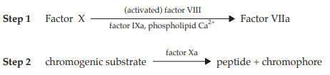
Fig. 1 Schematic Representation of the Assay of Human Coagulation Factor VIII
Both steps employ reagents that may be obtained commercially from a variety of sources. Although the composition of individual reagents may be subject to some variation, their essential features are described in the following specification. Deviations from this description may be permissible provided that it has been shown, using the International Standard for Human Blood Coagulation Factor VIII Concentrate as the standard, that the results obtained do not differ significantly.
It is important to demonstrate by validation the suitability of the kit used, notably by checking the time course of factor Xa generation in order to determine the time taken to reach 50 per cent of the maximal factor Xa generation.
REAGENTS
The coagulation factor reagent comprises purified proteins derived from human or bovine sources. These include factor X, factor IXa, and a factor VIII activator, usually thrombin. These proteins are partly purified, preferably to at least 50 per cent, and do not contain impurities that interfere with the activation of factor VIII or factor X. Thrombin may be present in its precursor form prothrombin, provided that its activation in the reagent is sufficiently rapid to give almost instantaneous activation of factor VIII in the assay. Phospholipid may be obtained from natural sources or be synthetically prepared, and must, to a substantial extent, consist of the species phosphatidylserine. The components of the complete reagent are usually divided into at least two separate reagents, each lacking the ability to generate factor Xa on its own. One of the reagents contains calcium ions. After reconstitution, the reagents may be combined provided that no substantial amounts of factor Xa are generated in the absence of factor VIII. In the final incubation mixture, factor VIII must be the only rate-limiting component.
The second step comprises the quantification of the formed factor Xa, employing a chromogenic substrate that is specific for factor Xa. Generally this consists of a derivatized short peptide of between 3 and 5 amino acids, joined to a chromophore group. On cleavage of this group from the peptide substrate, its chromophoric properties shift to a wavelength allowing its spectrophotometric quantification. The substrate must also contain appropriate inhibitors to stop further factor Xa generation, e.g. chelating agents, and to suppress thrombin activity.
ASSAY
Reconstitute the entire contents of one ampoule of the reference preparation and the preparation to be examined; use immediately. Add sufficient prediluent to the reconstituted preparations to produce solutions containing 0.5 to 2.0 IU per ml.
The prediluent consists of hemophilia A plasma, or of an artificially prepared reagent that contains sufficient von Willebrand factor and that give results that do not differ significantly from those obtained employing hemophilia plasma.The prediluted materials must be stable beyond the time required for the assay.
Prepare further dilutions of reference and test preparations using non-chelating, appropriately buffered solution, for example, tris(hydroxymethyl)methylamine or imidazole, containing 1 per cent of bovine serum albumin or human albumin. Prepare at least two dilution series of at least three further dilutions for each material. Prepare the dilutions such that the final factor VIII concentration in the reaction mixture is preferably below 0.01 IU per ml, during the step of factor Xa generation.
Prepare a control solution that includes all components except factor VIII.
Prepare all dilutions in plastic tubes and use immediately.
Step 1 Mix prewarmed dilutions of the factor VIII reference preparation and of the preparation to be examined with an appropriate volume of the prewarmed coagulation factor reagent or a combination of its separate constituents, and incubate the mixture in plastic tubes or microplate wells at 37º. Allow the activation of factor X to proceed for a suitable time, terminating the reaction (step 2) when the factor Xa concentration has reached approximately 50 per cent of the maximal (plateau) level. Appropriate activation times are usually between 2 and 5 minutes.
Step 2 Terminate the activation by addition of a prewarmed reagent containing a chromogenic substrate. Quantify the rate of substrate cleavage, which must be linear with the concentration of factor Xa formed, by measuring the absorbance change at an appropriate wavelength using a spectrophotometer, either monitoring the absorbance continuously, thus allowing the initial rate of substrate cleavage to be calculated, or terminating the hydrolysis reaction after a suitable interval by lowering the pH by addition of a suitable reagent, such as a 50 per cent v/v solution of acetic acid, or a 1 M citrate buffer solution pH 3. Adjust the hydrolysis time to achieve a linear development of chromophore over time. Appropriate hydrolysis times are usually between 3 and 15 minutes, but deviations are permissible if better linearity of the dose-response relationship is thus obtained.
Calculate the potency of the test preparation as described in the “Statistical Analysis of Results of Biological Assays and Tests” (Appendix 9).
15.1.4 Biological Assay of Human Coagulation Factor IX
The principle of the assay is to measure the ability of a factor IX preparation to reduce the prolonged coagulation time of factor IX-deficient plasma. The reaction is accelerated by addition of a reagent containing phospholipid and a contact activator, e.g. kaolin, silica or ellagic acid. The potency is assessed by comparing the dose-response curve of the preparation to be examined to that of a reference preparation, calibrated in International Units.
Reconstitute separately the preparation being examined and the reference preparation as stated on the label and use immediately. Where applicable, determine the amount of heparin present, (Appendix 14.2.1), and neutralize the heparin, for example by addition of protamine sulfate (10 μg of protamine sulfate neutralizes 1 IU of heparin). Predilute the preparation being examined and the reference preparation in factor IX-deficient plasma (for example plasma substrate 2) to produce solution containing 0.5 to 2.0 IU per ml. Prepare at least three dilutions for each material, preferably in duplicate, using a suitable buffer solution (for example imidazole buffer solution pH 7.3) containing 1 per cent w/v solution of bovine serum albumin or human albumin.
Use this dilutions immediately. Use an apparatus suitable for measurement of coagulation times or carry out the assay with incubation tubes maintained in a water-bath at 37º. Place in each tube 0.1 ml of factor IX-deficient plasma (for example plasma substrate 2) and 0.1 ml of one of the dilutions of the reference preparation or of the preparation to be examined. Add to each tube 0.1 ml of a suitable Activated Partial Thromboplastin Time (APTT) reagent containing phospholipid and contact activator and incubate the mixture for a recommended time at 37º. To each tube, add 0.1 ml of a 0.37 per cent w/v solution of calcium chloride previously heated to 37º. Using a timer, measure the coagulation time, i.e. the interval between the moment of the addition of the calcium chloride and the first indication of the formation of fibrin. The volumes given above may be adapted to the APTT reagent and apparatus used. Calculate the potency of the test preparation as described in the “Statistical Analysis of Results of Biological Assays and Tests (Appendix 9).
15.1.5 Biological Assay of Human Coagulation Factor X
Human coagulation factor X is assayed following specific activation to form factor Xa. Factor Xa is estimated by comparing its activity in cleaving a specific chromogenic peptide substrate with the same activity of the International Standard or of a reference preparation calibrated in International Units.
The chromogenic assay method consists of two steps: snake venom-dependent activation of factor X, followed by enzymatic cleavage of a chromogenic factor Xa substrate to form a chromophore that can be quantified spectrophotometrically. Under appropriate assay conditions, there is a linear relation between factor Xa activity and the cleavage of the chromogenic substrate.
REAGENTS
Russell’s viper venom specific factor X activator (RVV) A protein derived from the venom of Russell’s viper (Vipera russelli) which specifically activates factor X. Reconstitute according to the manufacturer’s instructions. Store the reconstituted preparation at 4º and use within 1 month.
Factor Xa chromogenic substrate Specific chromogenic substrate for factor Xa such as: N-α benzyloxycarbonyl-D-arginyl-L-glycyl-L-arginine-4- nitroanilide dihydrochloride, N-benzoyl-L-isoleucyl-Lglutamyl-glycyl-L-arginine-4-nitroanilide hydrochloride, methanesulfonyl-D-leucyl-glycyl-L-arginine-4- nitroanilide, methoxycarbonyl-D-cyclohexylalanylglycyl-L-arginine-4-nitroanilide acetate. Reconstitute according to the manufacturer’s instructions.
Dilution buffer Solution containing 0.37 per cent w/v of tris(hydroxymethyl)methylamine, 1.8 per cent w/v of sodium chloride, 0.21 per cent w/v of imidazole, 0.002 per cent w/v of hexadimethrine bromide and 0.1 per cent w/v of bovine serum albumin or human albumin. Adjust to pH 8.4 if necessary, using hydrochloric acid.
METHOD
Test solution Dilute the preparation being examined with dilution buffer to obtain a solution containing 0.18 IU of factor X per ml. Prepare at least three further dilutions in dilution buffer.
Reference solution Dilute the reference preparation to be examined with dilution buffer to obtain a solution containing 0.18 IU of factor X per ml. Prepare at least three further dilutions in dilution buffer.
Warm all solutions to 37º in a water-bath shortly before the test.
The following working conditions apply to microtitre plates. If the assay is carried out in tubes, the volumes are adjusted while maintaining the proportions in the mixture.
Using a microtitre plate maintained at 37º, add 12.5 μl of each dilution of the test solution or the reference solution to each of a series of wells. To each well add 25 μl of RVV and incubate for exactly 90 seconds. To each well add 150 μl of factor X chromogenic substrate, diluted 1 in 6 in dilution buffer.
Read the rate of change of absorbance at 405 nm (Appendix 2.2) continuously over a period of 3 minutes and obtain the mean rate of change of absorbance (ΔA/minute). If continuous monitoring is not possible, read the absorbance at 405 nm at suitable consecutive intervals, for instance 40 seconds, plot the absorbances against time on a linear graph and calculate ΔA/minute as the slope of the line. From the ΔA/minute values of each individual dilution of standard and test preparations, calculate the potency of the preparation being examined and check the validity of the assay by the “Statistical Analysis of Results of Biological Assay and Tests” (Appendix 9).
15.1.6 Biological Assay of Anti-Rh0 (D) Immunoglobulin
Method A
The potency of anti-Rh0 (D) immunoglobulin is determined by comparing the quantity necessary to produce agglutination of Rh0 (D)-positive red blood cells with the quantity of a reference preparation, calibrated in International Units, required to produce the same effect.
Use pooled Rh0 (D)-positive red blood cells, collected not more than 7 days earlier and suitably stored, obtained from not less than four group O R1R1 donors. To a suitable volume of the cells, previously washed three times with saline TS, add an equal volume of bromelains TS, allow to stand at 37º for 10 minutes, centrifuge, remove the supernatant liquid and wash three times with saline TS. Suspend 20 volumes of the red blood cells in a mixture of 15 volumes of inert serum, 20 volumes of a 30 per cent w/v solution of bovine serum albumin and 45 volumes of saline TS. Stand the resulting suspension in iced water, stirring continuously.
Using a calibrated automated dilutor, prepare suitable dilutions of the preparation being examined and of the reference preparation using as diluent a solution containing 0.5 per cent w/v of bovine serum albumin in saline TS.
Use a suitable apparatus for automatic continuous analysis. The following protocol is usually suitable: maintain the temperature in the manifold, except for the incubation coils, at 15.0º. Pump into the manifold of the apparatus the red blood cell suspension at a rate of 0.1 ml per minute and a 0.3 per cent w/v solution of methylcellulose 450 at a rate of 0.05 ml per minute. Introduce the dilutions of the preparation being examined and the reference preparation at a rate of 0.1 ml per minute for 2 minutes, followed by the diluent solution at a rate of 0.1 ml per minute for 4 minutes before the next dilution is introduced.
Introduce air at a rate of 0.6 ml per minute. Incubate at 37º for 18 minutes and then disperse the rouleaux1 by introducing at a rate of 1.6 ml per minute saline TS containing a suitable wetting agent (for example, polysorbate 20 at a final concentration of 0.02 per cent w/v) to prevent disruption of the bubble pattern. Allow the agglutinates to settle and decant twice, first at 0.4 ml per minute and then at 0.6 ml per minute. Lyse the unagglutinated red blood cells with a solution containing 0.5 per cent w/v of octoxinol 10, 0.02 per cent w/v of potassium hexacyanoferrate(III), 0.1 per cent w/v of sodium hydrogencarbonate and 0.005 per cent w/v of potassium cyanide at a rate of 2.5 ml per minute. A tenminute delay coil is introduced to allow for conversion
1 An abnormal group of red blood cells, adhering together like a roll of coins.
of the hemoglobin. Continuously record the absorbance (Appendix 2.2), of the hemolysate at a wavelength between 540 and 550 nm. Determine the range of antibody concentrations over which there is a linear relationship between concentration and the resultant change in absorbance (ΔA). From the results, prepare a standard curve and use the linear portion of the curve to determine the activity of the preparation being examined.
Calculate the potency of the preparation to be examined using the “Statistical Analysis of Results of Biological Assay and Test” (Appendix 9).
Method B
The potency of anti-Rh0 (D) immunoglobulin is determined by competitive enzyme-linked immunoassay on erythrocyte-coated microtitre plates. The method is based on the competitive binding between a polyclonal anti-Rh0 (D) immunoglobulin preparation and a biotinylated monoclonal anti-Rh0 (D) antibody directed against a D-antigen specific epitope. The activity of the preparation to be examined is compared with a reference preparation calibrated in International Units.
MATERIALS
Reagents not specified are of analytical grade.
Phosphate-buffered saline (PBS) Dissolve 8.0 g of sodium chloride, 0.76 g of anhydrous disodium hydrogenphosphate, 0.2 g of potassium chloride, 0.2 g of potassium dihydrogenphosphate and 0.2 g of sodium azide in water and dilute to 1000 ml with the same solvent.
Tris-buffered saline (TBS) Dissolve 8.0 g of sodium chloride and 0.6 g of tris(hydroxymethyl)methylamine in water. Adjust to pH 7.2 (Appendix 4.11) with 1 M hydrochloric acid and dilute to 1000 ml with the same solvent.
Papain solution Prepare a solution by stirring 1 g of papain at 37º for 30 minutes in 10 ml of 0.067 M phosphate buffer solution pH 5.4, centrifuge at 10,000 × g for 5 minutes and filter through a membrane with a pore size of 0.22 μm. To activate, combine 1 ml of the filtrate with 1 ml of a 4.844 per cent w/v solution of Lcysteine and 1 ml of a 0.372 per cent w/v solution of sodium edetate and dilute to 10 ml with 0.067 M phosphate buffer solution pH 5.4. Freeze in aliquots at –20º or below.
Red blood cells Use pooled Rh0 (D)-positive red blood cells obtained from not less than three group O R2R2 donors. Wash the cells four times with PBS. Centrifuge the cells at 1800 × g for 5 minutes, mix a suitable volume of prewarmed packed cells with a suitable volume of prewarmed papain solution (2 volumes to 1 volume has been found suitable) and incubate at 37º for 10 minutes. Wash the cells four times with PBS. Store at 4º in an appropriate stabilizer for up to 1 week.
Biotinylated Brad-5 Use according to instructions.
Alkaline phosphatase-conjugated avidin streptavidin reagent Preferably modified to combine high specific activity with low non-specific binding. Use according to instructions.
Substrate solution Use para-nitrophenyl phosphate according to instructions.
Cell fixation buffer Dissolve 18.02 g of dextroxe, 4.09 g of sodium chloride, 1.24 g of boric acid, 10.29 g of sodium citrate and 0.74 g of sodium edetate in water. Adjust to pH 7.2 to 7.3 (Appendix 4.11) using 1 M sodium hydroxide or 1 M hydrochloric acid, dilute to 1000 ml with water. Use directly from storage at 4º.
Glutaraldehyde solution Immediately before use, add 90 μl of a 25 per cent w/v solution of glutaraldehyde to 24 ml of cold PBS.
Microtitre plates Plates to be coated with red blood cells are flat-bottomed polystyrene plates with surface properties optimized for enzyme immunoassay and high protein-binding capacity. Plates used to prepare immunoglobulin dilutions are U or V-bottomed polystyrene or polyvinyl chloride plates.
METHOD
Prepare a 0.1 per cent v/v suspension of papaintreated red blood cells in cold cell fixation buffer. Pipette 50 μl into each well of the flat-bottomed microtitre plate.
Centrifuge the plate at 350 × g for 3 minutes, preferably at 4º. Without removing the supernatant, gently add 100 μl of glutaraldehyde solution to each well and leave for 10 minutes. Drain the wells by quickly inverting the plate and wash three times with 250 to 300 μl of PBS. This may be done manually or using a suitable automated plate washer. Either carry out the assay as described below, or store the plate at 4º after draining off the PBS and adding 100 μl of cell fixation buffer per well and sealing with plastic film. Plates can be stored at 4º for up to 1 month.
Test solutions For freeze-dried preparations, reconstitute as stated on the label. Prepare four independent replicates of five serial twofold dilutions starting with 30 IU per ml in PBS containing 1 per cent w/v of bovine serum albumin. If necessary, adjust the starting dilution to obtain responses falling in the linear portion of the dose-response curve.
Reference solutions Reconstitute the reference preparation according to instructions. Prepare four independent replicates of five serial twofold dilutions starting with 30 IU per ml in PBS containing 1 per cent w/v of bovine serum albumin.
Using U or V-bottomed microtitre plates, add 35 μl of each of the dilutions of the test solution or reference solution to each of a series of wells. To each well add 35 μl of biotinylated Brad-5 at 250 ng per ml.
Empty the wells of the red cell-coated plate by inverting and draining on a paper towel. Add 250 μl of PBS containing 2 per cent w/v of bovine serum albumin and leave at room temperature for 30 minutes.
Empty the wells of the red cell-coated plate by inverting and draining on a paper towel and transfer 50 μl from each of the dilutions of the test solution or reference solution containing biotinylated Brad-5 into the wells. Use 50 μl of PBS containing 1 per cent w/v of bovine serum albumin as negative control. Seal the plate with plastic film and incubate at room temperature for 1 hour.
Remove liquid from the wells of the red cell-coated plate and wash three times with 250 to 300 μl of TBS.
Dilute the alkaline phosphatase-conjugated avidin/ streptavidin reagent in TBS containing 1 per cent w/v solution of bovine serum albumin and add 50 μl to each well. Incubate for 30 minutes at room temperature. Remove liquid from the wells of the red cell-coated plate and wash three times with 250 to 300 μl of TBS.
Add 100 μl of substrate solution to each of the wells and incubate at room temperature for 10 minutes in the dark. To stop the reaction, add 50 μl of 3 M sodium hydroxide to each of the wells.
Measure the absorbances at 405 nm and substract the negative control reading. Use the absorbance values in the linear range of the titration curve to estimate the potency of the preparation to be examined by the “Statistical Analysis of Results of Biological Assays and Tests” (Appendix 9).
Method C
The potency of anti-Rh0 (D) immunoglobulin is determined by flow cytometry in a microtitre plate format. The method is based on the specific binding between anti-Rh0 (D) immunoglobulin and Rh0 (D)- positive red blood cells. The activity of the preparation to be examined is compared with a reference preparation calibrated in International Units.