ตำรายาของประเทศไทย
Thai Pharmacopoeia
สำนักยาและวัตถุเสพติด กรมวิทยาศาสตร์การแพทย์ กระทรวงสาธารณสุข
Bureau of Drug and Narcotic, Department of Medical Sciences, Ministry of Public Healthสำนักยาและวัตถุเสพติด กรมวิทยาศาสตร์การแพทย์ กระทรวงสาธารณสุข
Bureau of Drug and Narcotic, Department of Medical Sciences, Ministry of Public HealthAPPENDIX 14 BIOLOGICAL AND BIOCHEMICAL ASSAYS
14.2 BLOOD AND RELATED PRODUCTS
14.2.1 Biological Assay of Heparin in Coagulation Factors
Heparin is assayed as a complex with antithrombin III (AT) via its inhibition of coagulation factor Xa (antiXa activity). An excess of AT is maintained in the reaction mixture to ensure a constant concentration of the heparin-AT complex. Factor Xa is neutralized by the heparin-AT complex and the residual factor Xa hydrolyses, a specific chromogenic peptide substrate to release a chromophore. The quantity of chromophore is inversely proportional to the activity of the heparin.
Factor Xa chromogenic substrate Specific chromogenic substrate for factor Xa such as: N-benzoyl-Lisoleucyl-L-glutamyl-glycyl-L-arginine-4-nitroanilide hydrochloride. Reconstitute according to the manufacturer’s instructions.
Dilution buffer A 0.605 per cent w/v solution of tris(hydroxymethyl)aminomethane. Adjust to pH 8.4 if necessary using hydrochloric acid.
Test solution Dilute the preparation to be examined with dilution buffer to obtain a solution expected to contain 0.1 IU of heparin per ml.
The following working conditions apply to microtitre plates. If the assay is carried out in tubes, the volumes are adjusted while maintaining the proportions in the mixture.
Warm all solutions to 37º in a water-bath shortly before the test.
Distribute in a series of wells, 20 μl of normal human plasma and 20 μl of antithrombin III solution 1. Add to the wells a series of volumes (20 μl, 60 μl, 100 μl and 140 μl) of the test solution or the reference solution and make up the volume in each well to 200 μl using dilution buffer (0.02 to 0.08 IU of heparin per ml in the final reaction mixture).
End-point method Transfer 40 μl from each well to a second series of wells, add 20 μl of bovine factor Xa solution and incubate at 37º for 30 seconds. Add 40 μl of a 1 mmol per litre solution of factor Xa chromogenic substrate and incubate at 37º for 3 minutes. Terminate the reaction by lowering the pH by the addition of a suitable reagent, such as a 20 per cent v/v solution of glacial acetic acid and measure the absorbance at 405 nm (Appendix 2.2). Appropriate reaction times are usually between 3 and 15 minutes, but deviations are permissible if better linearity of the dose-response relationship is thus obtained.
Kinetic method Transfer 40 μl from each well to a second series of wells, add 20 μl of bovine factor Xa solution and incubate at 37º for 30 seconds. Add 40 μl of a 2 mmol per litre solution of factor Xa chromogenic substrate, incubate at 37º and measure the rate of substrate cleavage by continuous measurement of the absorbance change at 405 nm (Appendix 2.2), thus allowing the initial rate of substrate cleavage to be calculated. This rate must be linear with the concentration of residual factor Xa.
Check the validity of the assay and calculate the heparin activity of the test preparation as described in the “Statistical Analysis of Results of Biological Assay and Tests” (Appendix 9, slope-ratio assay).
14.2.2 Test for Prekallikrein Activator
Prekallikrein activator (PKA) activates prekallikrein to kallikrein and may be assayed by its ability to cleave a chromophore from a synthetic peptide substrate so that the rate of cleavage can be measured spectrophotometrically and the concentration of PKA calculated by comparison with a standard preparation calibrated in International Units.
PREPARATION OF PREKALLIKREIN SUBSTRATE
(Note To avoid coagulation activation, blood or plasma used for the preparation of prekallikrein must come into contact only with plastics or silicone-treated glass surfaces.)
Draw 9 volumes of human blood into 1 volume of anticoagulant solution (ACD, CPD or 3.8 per cent w/v solution of sodium citrate) to which 1 mg per ml of hexadimethrine bromide has been added. Centrifuge the mixture at 3600 × g for 5 minutes. Separate the plasma and centrifuge again at 6000 × g for 20 minutes to sediment platelets. Separate the platelet-poor plasma and dialyse against 10 volumes of buffer A for 20 hours. Apply the dialyzed plasma to a chromatography column containing agarose -DEAE for ion exchange chromatography which has been equilibrated in buffer A and is equal to twice the volume of the plasma. Elute from the column with buffer A at about 20 ml per cm2 per hour. Collect the eluate in fractions and record the absorbance at 280 nm (Appendix 2.2). Pool the fractions containing the first protein peak so that the volume of the pool is about 1.2 times the volume of the platelet-poor plasma.
Test the substrate pool for absence of kallikrein activity by mixing 1 part with 20 parts of the prewarmed chromogenic substrate solution to be used in the assay and incubate at 37º for 2 minutes. The substrate is suitable if the increase in absorption is less than 0.001 per minute. Add 0.7 per cent w/v solution of sodium chloride to the pooled and filter using a membrane filter (porosity 0.45 μm). Freeze the filtrate in portions and store at –25º; the substrate may be freezedried before storage.
Carry out all procedures from the beginning of the chromatography to freezing in portions during a single working day.
METHOD
The assay may be carried out using an automated enzyme analyzer or a suitable microtitre plate system allowing kinetic measurements, with appropriate software for calculation of results. Standards, samples and prekallikrein substrate may be diluted as necessary using buffer B.
Incubate diluted standards or samples with prekallikrein substrate for 10 minutes such that the volume of the undiluted sample does not exceed 1/10 of the total volume of the incubation mixture to avoid errors caused by variation in ionic strength and pH in the incubation mixture. Incubate the mixture or a part thereof with at least an equal volume of a solution of a suitable synthetic chromogenic substrate, known to be specific for kallikrein (for example, N-benzoyl-L-prolylL-phenylalanyl-L-arginine 4-nitroanilide acetate or Dprolyl-L-phenylalanyl-L-arginine-4-nitroanilide dihydrochloride), dissolved in buffer B. Record the rate of change in absorbance per minute for 2 to 10 minutes at the wavelength specific for the substrate used. Prepare a blank for each mixture of sample or standard using buffer B instead of prekallikrein substrate.
Correct ΔA per minute by subtracting the value obtained for the corresponding blank. Plot a calibration curve using the values thus obtained for the reference preparation and the respective concentrations. Use the curve to determine the PKA activity of the preparation being examined.
REAGENTS
| Buffer A | |
| Tris(hydroxymethyl)methylamine | 6.055 g |
| Sodium chloride | 1.17 g |
| Hexadimethrine bromide | 0.050 g |
| Sodium azide | 0.100 g |
Dissolve the ingredients in water, adjust to pH 8.0 with 2 M hydrochloric acid and dilute to 1000 ml with water.
| Buffer B | |
| Tris(hydroxymethyl)methylamine | 6.055 g |
| Sodium chloride | 8.77 g |
Dissolve the ingredients in water, adjust to pH 8.0 with 2 M hydrochloric acid and dilute to 1000 ml with water.
14.2.3 Determination of Hemoglobin Concentration by Hemiglobincyanide Method
There are three methods for estimation of hemoglobin concentration by using a spectrometer or photoelectric colourimeter, i.e. alkaline-hematin, oxyhemoglobin (HbO2) and hemiglobincyanide or cyanomethemoglobin (HiCN) methods. A major advantage of the hemiglobincyanide (HiCN) method is the availability of a stable and reliable reference solution.
The hemiglobincyanide (HiCN) method is provided for determination of hemoglobin concentration in human blood. The basis of the method is dilution of blood in the solution containing potassium cyanide and potassium hexacyanoferrate(III). Hemoglobin (Hb) and its derivatives, methemoglobin (Hi) and carboxyhemoglobin (HbCO), but not sulphemoglobin (SHb) are oxidized to methemoglobin in the presence of alkaline potassium hexacyanoferrate(III). Methemoglobin then reacts with potassium cyanide to form hemiglobincyanide (HiCN) which has maximum absorption at 540 nm. The colour intensity measured at 540 nm is proportional to the total hemoglobin concentration.
CYANIDE-HEXACYANOFERRATE(III) SOLUTION
| Potassium hexacyanoferrate(III) | 200 mg |
| Potassium cyanide1 | 50 mg |
| Potassium dihydrogenphosphate | 140 mg |
| Non-ionic detergent2 | 0.5 ml |
| Water to | 1000 ml |
The solution should be clear and pale yellow in colour. When measured against water as a blank in a spectrophotometer at a wavelenght of 540 nm, the optical density must read zero. The pH of the solution should lie between 7.0 and 7.4 and must be checked regularly, at least once a month. If stored between 4º and 25º in a brown borosilicate glass bottle, the solution will be kept for several months. It must not be allowed to freeze, as this can result in its decomposition and must be discarded if it becomes turbid, or if the pH is found to be outside the 7.0 to 7.4 range or if it has an absorbance other than zero at 540 nm against a water blank.
Instead of the solution described above, Drabkin’s cyanide-hexacyanoferrate(III) solution (Drabkin’s reagent) or similar products, some of which are available in powder or tablet form, are also used in clinical hemoglobinometry. It should, however, be borne in mind that with these solutions complete conversion of hemoglobin derivatives to HiCN takes about 20 minutes and that the resulting solution is often slightly turbid, giving results that are too high.
1Highly poisonous. Great care must be taken when handling this reagent and to ensure safe storage.
2A suitable non-ionic detergent, i.e. nonylphenylpolyethylene glycol or equivalent, may be used.
HEMIGLOBINCYANIDE REFERENCE SOLUTION
Solutions of HiCN are available commercially as reference solutions that conform to the international specifications. They contain 550 to 850 mg of hemoglobin per litre and the exact concentration is indicated on the label. The solution is dispensed in sealed appropriate containers. In use, the reference solution is regarded as a dilution of whole blood, and the original Hb that it represents is obtained by mutiplying the figure stated on the label by the dilution to be applied to the blood sample.
Blood sample The blood sample may be taken from a freely bleeding capillary puncture (finger, heel in infants) or may be collected by venesection. The capillary puncture must be deep enough to allow the blood to flow freely. A venous specimen may be collected into any solid anticoagulant (EDTA, mixture of ammonium and potassium oxalate or heparin). The blood should be well mixed before sampling, either by gentle inversion at least 20 times, or, preferably, on a mechanical mixing device for 2 to 3 minutes.
Procedure Dilute 20.0 μl of the blood sample in 5.0 ml of Cyanide-hexacyanoferrate(III) solution. Mix well and allow to stand for at least 3 minutes to ensure the completion of the reaction. Measure the absorbances of Hemiglobincyanide reference solution and blood sample at the maximum at about 540 nm (Appendix 2.2), against Cyanide-hexacyanoferrate(III) solution as a blank.
Determine total hemoglobin concentration (g/dl) of blood sample by the following formula:
Hb (g/dl) = (Au/As)(Cs)(Dilution factor/100)
in which Cs is the concentration, g/dl, of Hb in Hemiglobincyanide reference solution, and As and Au are the absorbances of Hemiglobincyanide reference solution and blood sample, respectively.
To prepare a calibration curve, set up series of five tubes. Pipette into these tubes the following amounts of Hemiglobincyanide Reference Solution.
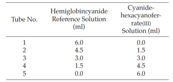
The hemoglobin concentration in the five tubes will be, respectively: labelled value (full strength); times labelled value;
 times labelled value;
 times labelled value; and zero strength (blank). Measure the absorbances of five tubes at the maximum at about 540 nm (Appendix 2.2), against Cyanide-hexacyanoferrate(III) solution as a blank. Construct a calibration curve by plotting the absorbance reading on the vertical axis and hemoglobin concentration on the horizontal axis of graphpaper. The points should fall on a straight line passing through zero. Determine total hemoglobin concentration (g/dl) of blood sample by reading from the calibration curve.